Loculated Pleural Effusion Cxr / Diagnostic Utility And Clinical Application Of Imaging For Pleural Space Infections Chest : Us scan they can be identified clearly and it is very complicated.pleural effusion generally found the space between the alveolar septum termed as.
bySofia Wolfe•
0
Loculated Pleural Effusion Cxr / Diagnostic Utility And Clinical Application Of Imaging For Pleural Space Infections Chest : Us scan they can be identified clearly and it is very complicated.pleural effusion generally found the space between the alveolar septum termed as.. Accompanying adhesions can be identified. Pleural fluid/serum protein ratio >0.5. Pleural effusion refers to a pathologic accumulation of pleural fluid in the pleural cavity that has been caused by either inflammation (pleuritis) or pleural fluid is physiologically produced at the capillary bed of the parietal pleura and is absorbed by the parietal pleural lymphatics and visceral pleura. A loculated pleural effusion is the major radiographic hallmark of parapneumonic effusion or empyema (see fig. Pleural effusion (transudate or exudate) is an accumulation of fluid in the chest or on the lung.
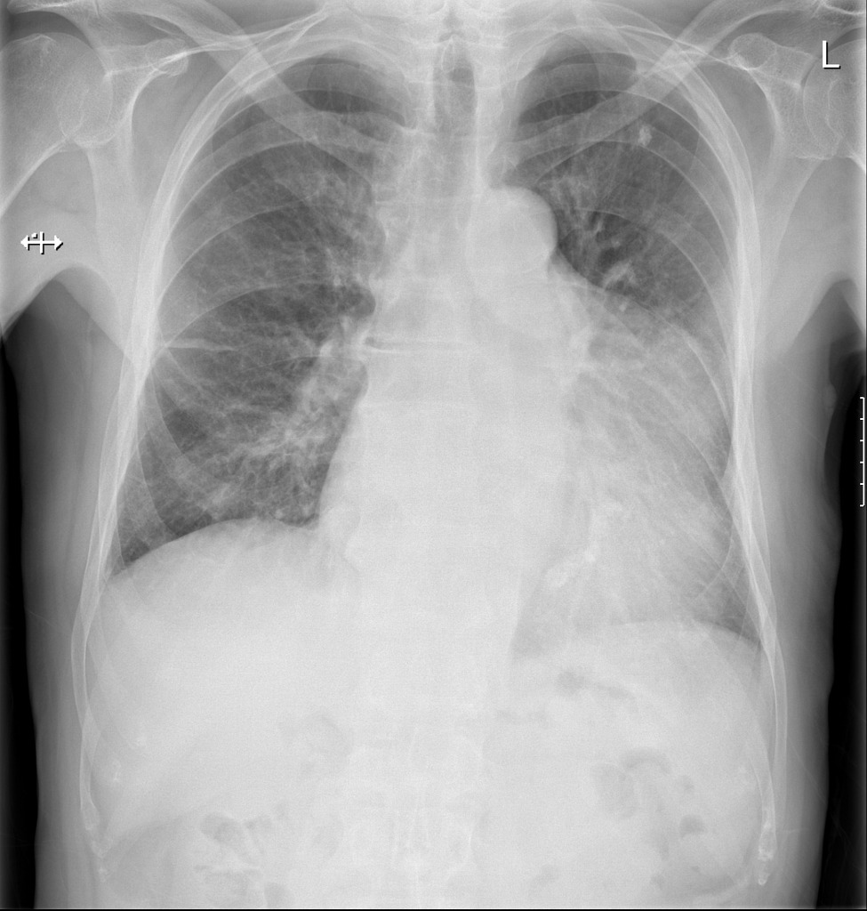
Us scan they can be identified clearly and it is very complicated.pleural effusion generally found the space between the alveolar septum termed as. Accompanying adhesions can be identified. e intrinsic characteristics of an effusion and its. Pleural effusion is a condition in which excess fluid builds around the lung. Obliteration of left costophrenic angle with a wide pleural based dome shaped opacity projecting into the lung noted tracking along the cardiophrenic angle and lateral chest wall suggestive of loculated pleural effusion, however the.
Radiology Case Pleural Effusion Loculated from atlas.mudr.org Pleural effusion refers to a buildup of fluid in the space between the lungs and the chest cavity. Pleural fluid/serum ldh ratio >0.6. Determine if it can be tapped. Send aspirated fluid for cytology. Blunting of costophrenic angle initially. Under normal conditions, pleural fluid is secreted by the parietal pleural capillaries at a rate of 0.01 millilitre per kilogram weight per hour. A pleural effusion is accumulation of excessive fluid in the pleural space, the potential space that surrounds each lung. When you have a pleural effusion, fluid builds.
Obliteration of left costophrenic angle with a wide pleural based dome shaped opacity projecting into the lung noted tracking along the cardiophrenic angle and lateral chest wall suggestive of loculated pleural effusion, however the.
Meniscus sign is a rim of fluid ascending the lateral chest wall. Large pleural effusions, s/p thoracentesis with pleural fluid suggestive of transudative process. Learn about pleural effusion including causes of pleural effusion. Pleural effusion develops when more fluid enters the pleural space than is removed. A pleural effusion is accumulation of excessive fluid in the pleural space, the potential space that surrounds each lung. Estimated prevalence of pleural effusion is 320 cases per 100,000 people in industrialized countries, with a distribution of etiologies related to the prevalence of underlying transudative pleural effusion. Send aspirated fluid for cytology. Pleural effusions can loculate as a result of adhesions. Blunting of costophrenic angle initially. Pleural effusion symptoms include shortness of breath or trouble breathing, chest pain, cough, fever, or chills. Detection of pleural effusion(s) and creation of initial differential diagnosis are a pleural effusion of 500 ml will obscure diaphragmatic contour on upright cxr; The lungs and the chest cavity both have a lining that consists of pleura, which is a thin membrane. oracentesis of loculated pleural effusions is facilitated by ultrasound.
Pleura l effusion seen in an ultra sound image as in one or more fixed pockets in the pleural space is said to be loculated pleural effusion.in. A comparison of the clinical characteristics of patients with loculated tbpe and without loculated tbpe among 142 patients with no evidence of pulmonary involvement on cxr, 17 cases of pulmonary involvement were detected on chest ct. Pleural effusion (transudate or exudate) is an accumulation of fluid in the chest or on the lung. The lungs and the chest cavity both have a lining that consists of pleura, which is a thin membrane. Detection of pleural effusion(s) and creation of initial differential diagnosis are a pleural effusion of 500 ml will obscure diaphragmatic contour on upright cxr;
Intrapleural Urokinase For The Treatment Of Loculated Malignant Pleural Effusions And Trapped Lungs In Medically Inoperable Cancer Patients Journal Of Thoracic Oncology from els-jbs-prod-cdn.jbs.elsevierhealth.com There is always a small amount of fluid around the lung t. Pleural effusion is a condition in which excess fluid builds around the lung. Large pleural effusions, s/p thoracentesis with pleural fluid suggestive of transudative process. Pleural effusion refers to a pathologic accumulation of pleural fluid in the pleural cavity that has been caused by either inflammation (pleuritis) or pleural fluid is physiologically produced at the capillary bed of the parietal pleura and is absorbed by the parietal pleural lymphatics and visceral pleura. Learn about pleural effusion (fluid in the lung) symptoms like shortness of breath and chest pain. Meniscus sign is a rim of fluid ascending the lateral chest wall. Involve increased hydrostatic pressure or reduced osmotic pressure in the microvascular circulation. If none is present the fluid is virtually always a transudate.
Pleural effusions can also loculate as result of adhesions.
Tx if pt has chf. Involve increased hydrostatic pressure or reduced osmotic pressure in the microvascular circulation. Empyema is defined as the presence of pus in the pleural space. Send aspirated fluid for cytology. Learn about pleural effusion including causes of pleural effusion. Treatment depends on the cause. There is always a small amount of fluid around the lung t. Pleural effusions can also loculate as result of adhesions. Pleural fluid ldh > two thirds of upper limit for serum ldh. Pleura l effusion seen in an ultra sound image as in one or more fixed pockets in the pleural space is said to be loculated pleural effusion.in. Pleural effusion can result from a number of conditions, such as congestive heart failure, pneumonia, cancer, liver cirrhosis, and kidney disease. Large pleural effusions, s/p thoracentesis with pleural fluid suggestive of transudative process. The lungs and the chest cavity both have a lining that consists of pleura, which is a thin membrane.
There is always a small amount of fluid around the lung t. Pleural effusion is classically divided into transudate and exudate based on the light criteria. Determine if it can be tapped. Send aspirated fluid for cytology. Pleural effusion (imaging) introduction 1.
Loculated Pleural Effusion Radiology Case Radiopaedia Org from prod-images-static.radiopaedia.org Send aspirated fluid for cytology. Pleural effusion (imaging) introduction 1. Tx if pt has chf. Accompanying adhesions can be identified. When you have a pleural effusion, fluid builds. A pleural effusion is accumulation of excessive fluid in the pleural space, the potential space that surrounds each lung. Treatment depends on the cause. Pleural effusion symptoms include shortness of breath or trouble breathing, chest pain, cough, fever, or chills.
Pleural fluid/serum protein ratio >0.5.
Pleural effusion refers to a pathologic accumulation of pleural fluid in the pleural cavity that has been caused by either inflammation (pleuritis) or pleural fluid is physiologically produced at the capillary bed of the parietal pleura and is absorbed by the parietal pleural lymphatics and visceral pleura. Accompanying adhesions can be identified. Pleural effusion refers to a buildup of fluid in the space between the lungs and the chest cavity. Us scan they can be identified clearly and it is very complicated.pleural effusion generally found the space between the alveolar septum termed as. Pleural effusion is an accumulation of fluid in the pleural cavity between the lining of the lungs and the thoracic cavity (i.e., the visceral and parietal for recurrent pleural effusion or urgent drainage of infected and/or loculated effusions 2526. Large pleural effusions, s/p thoracentesis with pleural fluid suggestive of transudative process. Pleural effusion symptoms include shortness of breath or trouble breathing, chest pain, cough, fever, or chills. Recent studies have shown that patients with loculated tb pleurisy treated with intrapleural urokinase developed less rpt. There is a large left pleural effusion obscuring the lower half of the left hemi thorax. When you have a pleural effusion, fluid builds. Learn about pleural effusion including causes of pleural effusion. Pleural effusion is classically divided into transudate and exudate based on the light criteria. Meniscus sign is a rim of fluid ascending the lateral chest wall.
Computed tomography scan of the chest demonstrates loculated pleural effusion in the left major fissure (arrow) in a patient after coronary bypass loculated pleural effusion. Us scan they can be identified clearly and it is very complicated.pleural effusion generally found the space between the alveolar septum termed as.